I am on imaging this week and so far it has been fantastic (granted it is only Monday but it gives me high hopes for the days to come). Today we discussed a case that happened a few years ago concerning a Boston terrier. Boston terriers are predisposed to peritoneal-pericardial diaphragmatic hernia (PPDH). I know that is a huge scary word but it is really quite simple. Most people think of hernias in the abdomen (result of a weakening if the abdominal walls/muscles that allows a loop of intestines to slip out) and diaphragmatic hernias are very similar to that but it is when there is some sort of penetration of the diaphragm allowing the guts to slip through into the chest cavity. Now, PPDH is when the contents slip through the diaphragm and into the pericardial sac (the tissue covering the heart). This was actually an incidental finding, she came in due to concerns of dribbling urine. You would think that this would be a major problem, but it is actually a congenital issue found in some Boston terriers and they can live full lives with it. The main cause for surgical intervention is if something that expands were in the sac (i.e. stomach). The symptom for this would not be from the heart but from difficulty breathing due to pressure on lungs.
Now time for pictures, yay! All names and information has been removed for confidentiality.
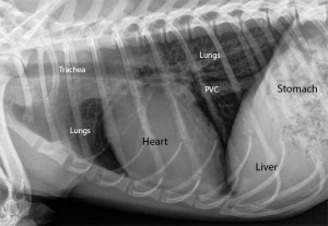
This image below shows a chest radiograph of what would be normal to see. The line separating the lungs and the liver/stomach would be the diaphragm. Since the patient is a Boston terrier, all of the organs will be slightly closer together but this gives you a good idea of what you should be seeing.
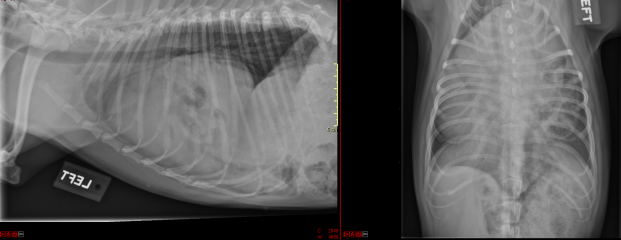
Now the images below are of the PPDH patient. You don’t have to be a radiograph-reading wiz to tell that something just isn’t right about this. The lung fields are small or missing (most of the lobes were actually collapsed), the heart is practically indiscernible, there are what appears to be loops where the heart is (shown by the darkened spaces which could be gas in the intestinal tract), and it is impossible to tell the entire length of where the diaphragm is.
So now you are probably wondering, or have forgotten more likely, about what happened with the urine dribbling issues. She was found to have bilateral ectopic ureters. The ureters are the tubes that bring the urine from the kidneys to the bladder and ectopic means that they are traveling to somewhere other than the bladder. This can be a not-too-uncommon cause of urinary incontinence in young dogs.
This pooch did have surgical intervention to restore all gut contents to their proper place and the ureters were placed back to their rightful place to the bladder.
That is all I have for you today but keep an eye out for future post where I discuss the bobcat my wildlife team is taking care of. I don’t want to make a post on it yet until we have a better idea of the prognosis and treatment timeline. We were also instructed to not post anything about it on the internet until future notice as it is a great PR opportunity that will garner a lot of attention and the managers wish to be the only ones currently putting out information so that it can be the most correct information. Hopefully we can get some funding in to help pay for the relatively expensive surgery it will need to be released into the wild.
Also, I am getting another dog. A ~4yr old great dane. This puts my pet count to 3 ferrets, 2 guinea pigs, and 3 dogs (I have issues haha). Here are some pictures of Buddy ft. Zella my border collie/lab mutt.


Neat case, did you get to see the surgery, or was it an old case? I thought I would love imaging rotation, but ended up hating it for reasons unrelated to imaging.
Also can I say, I’m jealous of your menagerie 🙂
LikeLike
Since I was on the imaging rotation I only got to see the imaging part of it. I thoroughly enjoyed my imaging rotation, it was fantastic. In the past few weeks, I had heard from nearly everyone who had the rotation that there was so much down-time that it was extremely boring. From my experience it was packed full of patients. I got to help with radiographs for dogs, cats, swine, and horses, fluoroscopy on a dog and a goat, ultrasounds on cats and dogs, nuclear scintigraphy on a horse, and CT’s on dogs and a potbelly pig. The very last day I actually got to practice my ultrasounding skills on a ball python.
Yeah I am on the brink of having too many pets haha. It is time to practice my self-control skills, however limited they may be.
LikeLike